1) electron probe X ray microanalysis


电镜X射线显微分析
1.
Three methods of specimen preparation, resin embedding section TEM specimen preparation, rapid frozen fixation cryoultrathin section and ethanolic phosphotungstic acid (EPTA) staining were applied to electron probe X ray microanalysis on the chromaffin granules in TEM.
通过比较常规透射电镜制样法、快速冷冻固定 冷冻超薄切片法及磷钨酸乙醇 (EPTA)染色法在嗜铬颗粒透射电镜X射线显微分析中的应用 ,发现磷钨酸乙醇染色法能使嗜铬颗粒电子着色 ,从而较好地显示嗜铬颗粒的超微结构。
2) Quantitative X-ray microanalysis


电镜X-射线显微定量分析
4) X-ray microanalyser


X光(X射线),显微分析器
5) electron microscopy and electron probe X-ray microanalzer


电镜及电子探针X射线微量分析
6) X-ray microscope


X射线显微镜
1.
A set of non-coaxial grazing X-ray microscope consisting of four spherical mirrors was designed for diagnosis of ICF,and the aberrations and imaging quality of the microscope were analyzed.
设计了一套由4个反射镜组成的非共轴掠入射X射线显微镜,分析了该系统的像差和成像质量。
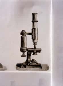
补充资料:显微镜19世纪中期的显微镜
[图]

说明:补充资料仅用于学习参考,请勿用于其它任何用途。
参考词条