1) shear force microscope


剪切力显微镜
1.
Contact mode atomic force microscope (AFM),friction force microscope (FFM),and shear force microscope (SFM) were used to map the patterns.
考察了膜的表面性质对接触式原子力显微镜(AFM),摩擦力显微镜(FFM)和剪切力显微镜(SFM)的成像的影响。
2) tuning-fork shear-force microscope(TSM)


调谐音叉剪切力显微镜(TSM)
3) light section microscope


光切显微镜
1.
The image extraction system of light section microscope is reformed.


改装了光切显微镜图像提取系统 ,采用数字摄像头摄取表面轮廓图像 ,通过图像处理 ,可提取被测表面的二维轮廓曲线 ,计算出多种表面粗糙度参数值 ,并可打印图形和相关报表。
2.
The paper presents a surface roughness measurement system which is based on light section principle and consists of conventional light section microscope and CCD camera as well as image processing software.
以光切法的测量原理为基础,应用传统双管光切显微镜、CCD摄像装置、虚拟仪器及图像处理技术开发了表面粗糙度检测系统。
4) Interphako interference microscopy


显微剪切干涉术
5) MFM


磁力显微镜
1.
Study on the effect of oblique sputtering on TbFe thin films by MFM;


利用磁力显微镜研究倾斜溅射对TbFe薄膜磁性能的影响
2.
Through the sample surface analysis by Atomic Force Microscope (AFM) and Magnetic Force Microscope (MFM), it is showed that the condition of annealing will effect the distribution of the grain in sample.
利用原子力显微镜和磁力显微镜对样品的表面进行分析,发现退火条件会影响样品磁性粒子的分布。
3.
In this paper, the using of Magnetic Force Microscopic (MFM) was introduced andthe transformations of magnetic domains were investigated by MFM.
本论文的工作主要分为两个方面:对磁力显微镜及其检测方法本身的研究和使用磁力显微镜研究薄膜磁畴结构的变化。
6) magnetic force microscopy


磁力显微镜
1.
It is analysed using magnetic force microscopy in this paper,and the result of magnetic force image ( MFI) shows that the sample induces a hard magnetic R_2M_(14)B(M=Fe or Co,R=Nd or Dy) phase after annealing.
5B20条带,运用磁力显微镜(MFM)的方法对该合金进行分析,从所得磁力图中看到试样退火后在基体上会析出一定量的硬磁相R2M14B(M=Fe或Co,R=Nd或Dy),其磁畴尺寸范围为200~500nm,磁畴尺寸远大于晶粒尺寸,磁畴跨越许多晶粒,即出现交换作用畴结构。
2.
The magnetic tips dependence of the magnetic domains in low coercivity magnetic (YGdBi)3(GaFe)_5O_12 garnet was investigated using magnetic force microscopy.
本文采用磁力显微镜(MFM)对磁性石榴石(YGdBi)3(GaFe)5O12薄膜的磁畴结构进行了观察研究。
3.
The magnetic tips dependence of the magnetic domains in low coercivity magnetic(YGdBi)_3(GaFe)_5O_(12) garnet was investigated using magnetic force microscopy.
本文采用磁力显微镜(MFM)对磁性石榴石(YGdBi)_3(GaFe)_5O_(12)薄膜的磁畴结构进行了观察研究。
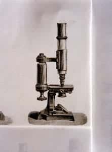
补充资料:显微镜19世纪中期的显微镜
[图]

说明:补充资料仅用于学习参考,请勿用于其它任何用途。
参考词条