1) magnetic resonance imaging


磁共振成像
1.
The physical principle of magnetic resonance imaging;


磁共振成像技术的物理学原理
2.
Application of magnetic resonance imaging in diagnosis and treatment of pelvic neoplasms;


磁共振成像在盆腔肿瘤诊治中的应用
3.
Analysis of magnetic resonance imaging in evaluating the myometrium invasion of endometrial carcinoma;
磁共振成像诊断子宫内膜癌肌层浸润深度
2) MRI
[英][,em ɑ:r 'aɪ] [美]['ɛm 'ɑr 'aɪ]


磁共振成像
1.
Progress in targeting MRI contrast agents;


靶向性磁共振成像造影剂的研究进展
2.
Clinical evaluation of neonatal hypoglycemic brain injury demonstrated by serial MRIs;


新生儿低血糖脑损伤临床特征与磁共振成像动态变化
3.
Analysis on Countermeasure for the Artifacts of MRI and Its Elimination;


磁共振成像的伪影及消除对策分析
3) MR imaging


磁共振成像
1.
Relevance of relative contrast enhanced index microvessel density and proliferating cell nuclear antigen in renal clear cell carcinoma with dynamic contrast enhanced MR imaging;
肾透明细胞癌磁共振成像动态增强相对强化指数与微血管密度、增殖细胞核抗原相关性探讨
2.
Methods Five patients(3 men,2 women;aged 21-39 years)with surgically and pathologically proved parathyroid adenoma underwent MR imaging of the parathyroid glands before the operation.
5T超导型磁共振成像系统。
3.
Objective To assess the diagnostic value of MR imaging of the wrist synovitis in early rheumatoid arthritis.
目的探讨磁共振成像(MRI)技术在早期类风湿关节炎(RA)患者腕关节滑膜病变临床诊断中的应用价值。
4) Magnetic resonance imaging(MRI)


磁共振成像
1.
Objective To study the gross anatomy of trigeminal nerves of rat,and to discuss the magnetic resonance imaging(MRI) characteristics of the nerves.
目的探讨大鼠三叉神经大体解剖形态及其磁共振成像(magnetic resonance imaging,MRI)方法和MRI表现。
2.
In the process of magnetic resonance imaging(MRI),the motion artifacts caused by patient s movement would degrade the image quality and make the detecting and locating the focus more complicated.
磁共振成像过程中,患者的轻微运动可产生运动伪影。
3.
Objective: To help radiologist look up magnetic resonance imaging(MRI)journals quickly and exactly, and to develop and use MRI sources from Medline database of NCBI.
目的 :为了方便放射学磁共振工作者快捷、准确地查阅有关核磁共振成像杂志文献 ,开发和利用 NCBI的 Medline数据库的核磁共振成像杂志文献资源。
5) magnetic resonance image


磁共振成像
1.
High field magnetic resonance image scanning of the normal ankle joint


正常踝关节的高场强磁共振成像扫描
2.
Objective To explore magnetic resonance image application value of knee joint injure.


目的探讨与总结磁共振成像在膝关节损伤的影像应用中的价值。
3.
Objective:To compare magnetic resonance image(MRI) and upper airway pressure monitoring for locating the site(s) of obstruction in the upper airway.
目的:研究磁共振成像(MRI)及上气道压力测定两种检查方法判定阻塞性睡眠呼吸暂停综合征(OSAS)患者阻塞平面的诊断符合率。
补充资料:磁共振成像
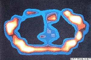
| 磁共振成像 magnetic resonance imaging 利用人体组织中某种原子核的核磁共振现象,将所得射频信号经过电子计算机处理,重建出人体某一层面的图像的诊断技术。又称核磁共振成像术。英文简称MRI。
MRI在临床上主要用于以下部位:①头部。可清晰分辨脑灰质和白质,对多发性硬化等一类脱髓鞘病优于CT。对脑外伤、脑出血、脑梗塞、脑肿瘤等同CT类似,但可显示CT为等密度的硬膜下血肿。脑梗塞或脑肿瘤的早期,CT不能查出,而MRI有可能显示。对钙化和脑膜瘤显示不好。脑干及小脑病变的MRI图像由于没有伪影是首选检查方法。②脊柱。不需要造影剂就能清晰区分脊髓、硬膜囊和硬膜外脂肪。对肿瘤、脊髓空洞症、脱髓鞘病变等均有较高诊断价值。显示骨折或脱位不如常规X射线或CT,但能观察脊髓损伤情况。显示椎间盘较好,可以分辨纤维环和髓核,特别是矢状面图像可以同时显示多个椎间盘突出。③四肢。对骨质本身病变显示不如常规X射线或CT。对软组织及肌肉病变包括肿瘤及炎症都能清晰显示,特别是对早期急性骨髓炎,是一种灵敏度很高的检查方法。也是检查膝关节半月板病变的首选方法。④盆腔。对直肠及泌尿生殖系统优于CT,无辐射损害,特别适用于孕妇及胎儿检查。⑤胸部。对肺的检查不如常规X射线,对纵隔检查则优于CT,不用造影剂即可分辨纵隔血管和肿物,也是一项有价值的心血管检查技术。⑥腹部。主要用于肝、胰、脾、肾等实质脏器。 |
说明:补充资料仅用于学习参考,请勿用于其它任何用途。
参考词条
